
تاريخ استخدام المجاهر الجراحية ودورها في جراحة الأعصاب
في تاريخ جراحة الأعصاب، تطبيقالمجاهر الجراحيةيمثل هذا رمزًا رائدًا، إذ ينتقل من العصر الجراحي العصبي التقليدي الذي كان يُجرى فيه الجراحة تحت العين المجردة إلى العصر الجراحي العصبي الحديث الذي يُجرى فيه الجراحة تحت...مجهرمن ومتى فعل ذلك؟مجاهر العمليات الجراحيةهل بدأ استخدامها في جراحة الأعصاب؟ ما هو دورهامجهر جراحيما هو الدور الذي لعبه في تطوير جراحة الأعصاب؟ مع تقدم العلوم والتكنولوجيا، هل سيلعب دورًا في ذلك؟مجهر جراحيهل يمكن استبدالها بمعدات أكثر تطوراً؟ هذا سؤال يجب على كل جراح أعصاب أن يكون على دراية به وأن يطبق أحدث التقنيات والأدوات في مجال جراحة الأعصاب، مما يعزز تحسين مهارات جراحة الأعصاب.
1- تاريخ تطبيقات المجهر في المجال الطبي
في الفيزياء، تُعرف عدسات النظارات بأنها عدسات محدبة ذات بنية واحدة لها تأثير تكبير، ويكون تكبيرها محدودًا، وتُعرف باسم العدسات المكبرة. في عام 1590، قام عالمان هولنديان بتركيب لوحين من العدسات المحدبة داخل أسطوانة رفيعة، وبذلك اخترعا أول جهاز تكبير ذي بنية مركبة في العالم:مجهربعد ذلك، جرى تحسين بنية المجهر باستمرار، وزاد التكبير بشكل متواصل. في ذلك الوقت، كان العلماء يستخدمون هذا بشكل أساسيمجهر مركبلمراقبة التراكيب الدقيقة للحيوانات والنباتات، مثل تركيب الخلايا. منذ منتصف القرن التاسع عشر وحتى أواخره، بدأ استخدام العدسات المكبرة والمجاهر تدريجيًا في المجال الطبي. في البداية، استخدم الجراحون عدسات مكبرة تشبه النظارات ذات عدسة واحدة، تُوضع على جسر الأنف لإجراء العمليات الجراحية. في عام 1876، أجرى الطبيب الألماني سايميش أول جراحة "مجهرية" في العالم باستخدام عدسة مكبرة مركبة تشبه النظارات (نوع الجراحة غير معروف). في عام 1893، اخترعت شركة زايس الألمانية...مجهر ثنائي العدسةتُستخدم بشكل أساسي للملاحظة التجريبية في المختبرات الطبية، وكذلك لمراقبة آفات القرنية والحجرة الأمامية في مجال طب العيون. في عام 1921، استنادًا إلى بحث مخبري حول تشريح الأذن الداخلية للحيوانات، استخدم طبيب الأنف والأذن والحنجرة السويدي نايلين جهازًا ثابتًامجهر جراحي أحادي العدسةقام بتصميم وتصنيع جهاز لإجراء جراحة التهاب الأذن الوسطى المزمن على البشر، وهي جراحة دقيقة حقيقية. بعد عام واحد، قدم الطبيب الأعلى رتبةً لنايلين، هلولمغرين، جهازًا آخر.مجهر جراحي ثنائي العدسةتم تصنيعها بواسطة شركة زايس في غرفة العمليات.
الأوائلمجاهر العمليات الجراحيةكان له العديد من العيوب، مثل ضعف الاستقرار الميكانيكي، وعدم القدرة على الحركة، وإضاءة محاور مختلفة، وتسخين العدسة الشيئية، وضيق مجال التكبير الجراحي، وما إلى ذلك. كل هذه الأسباب تحد من استخدامه على نطاق أوسع.المجاهر الجراحيةفي السنوات الثلاثين التالية، وبسبب التفاعل الإيجابي بين الجراحين ومصنعي الميكروسكوبأداءالمجاهر الجراحيةوقد تم تحسينه باستمرار،مجاهر جراحية ثنائية العدسة, مجاهر مثبتة على السطحتم تطوير عدسات التكبير، وإضاءة مصدر الضوء المحوري، والأذرع المفصلية التي يتم التحكم فيها إلكترونيًا أو بضغط الماء، والتحكم بدواسة القدم، وما إلى ذلك، تباعًا. في عام 1953، أنتجت شركة زايس الألمانية سلسلة من الكاميرات المتخصصةمجاهر جراحية لأمراض الأذنوهي مناسبة بشكل خاص للعمليات الجراحية على الآفات العميقة مثل الأذن الوسطى والعظم الصدغي. بينما أداءالمجاهر الجراحيةمع استمرار التحسن، تتغير عقلية الجراحين باستمرار. على سبيل المثال، نص الطبيبان الألمانيان زولنر وولشتاين على أنالمجاهر الجراحيةيجب استخدامها في جراحة تشكيل غشاء الطبل. منذ خمسينيات القرن الماضي، غيّر أطباء العيون تدريجيًا ممارسة استخدام المجاهر فقط في فحوصات العيون وأدخلوامجاهر جراحة الأذنفي جراحة العيون. ومنذ ذلك الحين،مجهر جراحيوقد تم استخدامها على نطاق واسع في مجالات طب الأذن وطب العيون.
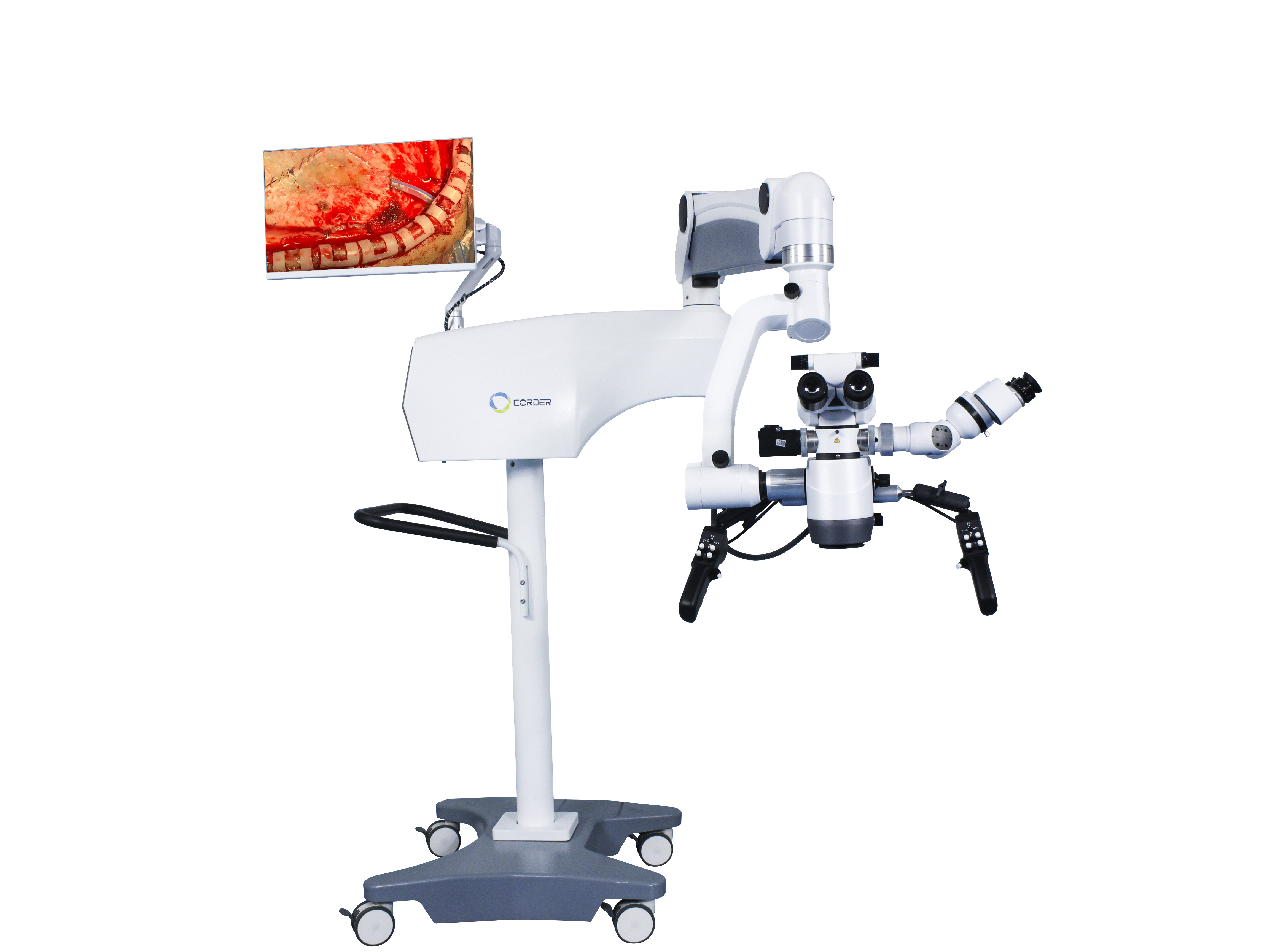
2- استخدام المجهر الجراحي في جراحة الأعصاب
نظراً لخصوصية جراحة الأعصاب، فإن تطبيقالمجاهر الجراحية في جراحة الأعصابيأتي هذا في وقت متأخر قليلاً عن طب الأذن وطب العيون، وكان جراحو الأعصاب يتعلمون هذه التقنية الجديدة بنشاط. في ذلك الوقت،استخدام المجاهر الجراحيةكان انتشاره الرئيسي في أوروبا. وقد قدمه طبيب العيون الأمريكي بيريت لأول مرة.المجاهر الجراحيةمن أوروبا إلى الولايات المتحدة في عام 1946، مما وضع الأساس الذي استخدمه جراحو الأعصاب الأمريكيونمجاهر العمليات الجراحية.
من منظور احترام قيمة الحياة البشرية، ينبغي أن تخضع أي تقنية أو معدات أو أدوات جديدة تُستخدم على جسم الإنسان لتجارب أولية على الحيوانات وتدريب تقني للمشغلين. في عام 1955، أجرى جراح الأعصاب الأمريكي ماليس جراحة دماغية على الحيوانات باستخداممجهر جراحي ثنائي العدسةأمضى كورتز، جراح الأعصاب في جامعة جنوب كاليفورنيا بالولايات المتحدة، عامًا كاملًا في تعلم التقنيات الجراحية باستخدام المجهر في المختبر بعد مشاهدة جراحة الأذن تحت المجهر. وفي أغسطس 1957، أجرى بنجاح جراحة ورم العصب السمعي لطفل يبلغ من العمر 5 سنوات باستخداممجهر جراحة الأذنوالتي كانت أول جراحة مجهرية في العالم. بعد ذلك بوقت قصير، نجح كورز في إجراء عملية توصيل العصب الوجهي بالعصب تحت اللسان للطفل باستخداممجهر جراحيوكان تعافي الطفل ممتازًا. وكانت هذه ثاني عملية جراحية دقيقة في العالم. وبعد ذلك، استخدم كورزه الشاحنات لنقله.مجاهر العمليات الجراحيةإلى أماكن مختلفة لإجراء جراحة الأعصاب المجهرية، وأوصى بشدة باستخدامالمجاهر الجراحيةإلى جراحي الأعصاب الآخرين. بعد ذلك، أجرى كورز عملية جراحية لربط تمدد الأوعية الدموية الدماغية باستخداممجهر جراحي(للأسف، لم ينشر أي مقالات). بدعم من مريضٍ كان يعالجه مصابًا بألم العصب ثلاثي التوائم، أسس أول مختبر في العالم لجراحة قاعدة الجمجمة المجهرية عام ١٩٦١. ينبغي لنا دائمًا أن نتذكر إسهامات كورزه في الجراحة المجهرية وأن نتعلم من شجاعته في تقبّل التقنيات والأفكار الجديدة. مع ذلك، وحتى أوائل التسعينيات، لم يتقبّل بعض جراحي الأعصاب في الصين هذه التقنيات.مجاهر جراحة الأعصابلإجراء العمليات الجراحية. لم تكن هذه مشكلة معمجهر جراحة الأعصابفي حد ذاتها، ولكن المشكلة تكمن في الفهم الأيديولوجي لجراحي الأعصاب.
في عام 1958، أسس جراح الأعصاب الأمريكي دوناغي أول مختبر في العالم لأبحاث وتدريب الجراحة المجهرية في برلينغتون، فيرمونت. في المراحل الأولى، واجه أيضًا ارتباكًا وصعوبات مالية من رؤسائه. في الأوساط الأكاديمية، كان يتصور دائمًا إمكانية فتح الأوعية الدموية القشرية لاستخراج الجلطات مباشرة من مرضى تجلط الدماغ. لذلك تعاون مع جراح الأوعية الدموية جاكوبسون في أبحاث على الحيوانات وأخرى سريرية. في ذلك الوقت، كان من الممكن خياطة الأوعية الدموية الصغيرة التي يبلغ قطرها 7-8 مليمترات أو أكثر بالعين المجردة. ولتحقيق مفاغرة طرفية للأوعية الدموية الدقيقة، حاول جاكوبسون أولًا استخدام عدسة مكبرة تشبه النظارات. بعد ذلك بوقت قصير، تذكر استخدامه لـمجهر جراحي لأمراض الأذن والأنف والحنجرةكان يعمل في مجال الجراحة عندما كان طبيباً مقيماً. لذلك، وبمساعدة شركة زايس في ألمانيا، صمم جاكوبسون مجهراً جراحياً ثنائي المشغل (منظار ثنائي القطب) لتوصيل الأوعية الدموية، مما يسمح لجراحين بإجراء الجراحة في وقت واحد. بعد تجارب مكثفة على الحيوانات، نشر جاكوبسون مقالًا عن التوصيل الجراحي المجهري للكلاب والشرايين غير السباتية (1960)، محققًا نسبة نجاح 100% في توصيل الأوعية الدموية. تُعد هذه الورقة البحثية الطبية رائدة في مجال جراحة الأعصاب المجهرية وجراحة الأوعية الدموية. كما صمم جاكوبسون العديد من الأدوات الجراحية المجهرية، مثل المقصات الدقيقة، وحوامل الإبر الدقيقة، ومقابض الأدوات الدقيقة. في عام 1960، أجرى دوناغي بنجاح عملية استئصال خثرة من شريان دماغي تحت إشراف طبي.مجهر جراحيبالنسبة لمريض مصاب بتجلط دماغي. بدأ روتون، من الولايات المتحدة، دراسة تشريح الدماغ تحت المجهر في عام 1967، رائدًا بذلك مجالًا جديدًا في علم التشريح الجراحي المجهري، ومساهمًا بشكل كبير في تطوير الجراحة المجهرية. نظرًا لمزاياالمجاهر الجراحيةومع تحسن أدوات الجراحة المجهرية، أصبح المزيد والمزيد من الجراحين يفضلون استخدامهاالمجاهر الجراحيةلإجراء العمليات الجراحية. ونشر العديد من المقالات ذات الصلة بإجراءات الجراحة المجهرية.
3- استخدام المجهر الجراحي في جراحة الأعصاب في الصين
بصفته صينيًا وطنيًا مغتربًا في اليابان، تبرع البروفيسور دو زيوي بأول مشروع محليمجهر جراحة الأعصابوما يتصل بهاأدوات الجراحة المجهريةالتحق بقسم جراحة الأعصاب في مستشفى كلية سوتشو الطبية (الذي أصبح الآن قسم جراحة الأعصاب في مستشفى جامعة سوتشو الأول) عام 1972. بعد عودته إلى الصين، أجرى في البداية عمليات جراحية دقيقة مثل تمدد الأوعية الدموية داخل الجمجمة والأورام السحائية. بعد أن علم بتوفرمجاهر جراحة الأعصابوباستخدام الأدوات الجراحية الدقيقة، زار البروفيسور تشاو يادو من قسم جراحة الأعصاب في مستشفى بكين ييوو البروفيسور دو زيوي من كلية سوتشو الطبية للاطلاع على كيفية استخدامالمجاهر الجراحيةقام البروفيسور شي يوكوان من مستشفى شنغهاي هواشان بزيارة شخصية لقسم البروفيسور دو زيوي لمشاهدة إجراءات الجراحة المجهرية. ونتيجة لذلك، انطلقت موجة من التعريف والتعلم والتطبيق لـمجاهر جراحة الأعصابوقد انطلقت هذه التقنية في مراكز جراحة الأعصاب الرئيسية في الصين، مما يمثل بداية جراحة الأعصاب الدقيقة في الصين.
4- تأثير الجراحة المجهرية
بسبب استخداممجاهر جراحة الأعصابتصبح العمليات الجراحية التي لا يمكن إجراؤها بالعين المجردة ممكنةً تحت ظروف التكبير من 6 إلى 10 أضعاف. على سبيل المثال، يمكن إجراء جراحة ورم الغدة النخامية عبر الجيب الغربالي لتحديد واستئصال أورام الغدة النخامية بأمان مع الحفاظ على الغدة النخامية السليمة؛ كما يمكن تحسين العمليات الجراحية التي لا يمكن إجراؤها بالعين المجردة، مثل أورام جذع الدماغ وأورام النخاع الشوكي. كان معدل الوفيات لدى الأكاديمي وانغ تشونغ تشنغ 10.7% في جراحة تمدد الأوعية الدموية الدماغية قبل استخدام هذه التقنية.مجهر جراحة الأعصاببعد استخدام المجهر في عام 1978، انخفض معدل الوفيات إلى 3.2%. معدل الوفيات لجراحة التشوه الشرياني الوريدي الدماغي بدون استخدام المجهرمجهر جراحيكانت النسبة 6.2%، وبعد عام 1984، مع استخداممجاهر جراحة الأعصابانخفض معدل الوفيات إلى 1.6%. استخداممجهر جراحة الأعصابيُتيح هذا الأسلوب علاج أورام الغدة النخامية عبر نهج جراحي طفيف التوغل عبر الأنف والوتدي دون الحاجة إلى فتح الجمجمة، مما يُقلل معدل الوفيات الجراحية من 4.7% إلى 0.9%. ويستحيل تحقيق هذه النتائج في ظل جراحة العيون التقليدية.المجاهر الجراحيةتُعدّ هذه الأدوات رمزاً لجراحة الأعصاب الحديثة، وقد أصبحت واحدة من المعدات الجراحية التي لا غنى عنها والتي لا يمكن الاستغناء عنها في جراحة الأعصاب الحديثة.
تاريخ النشر: 9 ديسمبر 2024


